Multi-function microplate inspection system with new imaging module and target recognition software
-Molecular Devices
Summary
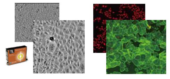
When it comes to obtaining high-quality, multi-parameter data, we need a sophisticated cytology-based instrument to increase data acquisition speed. Here we introduce a series of instruments based on cytology to introduce a cell imaging system and a multi-function microplate detection system, which can improve the throughput of detection and increase the richness of detection data. degree. The instrument is the latest SpectraMax® MiniMaxTM 300 from Molecular Devices, which comes standard with a white transmitted light channel and two fluorescence detection channels. Its patented dye-free cell detection technology allows accurate counting of unlabeled cells in a microplate through its white transmitted light channel, helping to analyze cell proliferation, differentiation, and the toxic effects of compounds on cells. Its standard two fluorescent channels greatly enhance the detection range of this instrument, including detection of cell proliferation, transfection efficiency, cell viability / apoptosis, cell differentiation or cell cycle. The use of new logic algorithms to identify cells in the analysis software simplifies the analysis process and outputs more parameter data: cell number, cell confluence, mean and intact cell area, average and intact fluorescence signal intensity. We cite several examples based on cell dynamics assays. We show how to use the new cell imaging system to better serve the research of biology. Based on this platform, we have developed several detection methods suitable for biopharmaceutical companies as well as research centers.
SpectraMax® MiniMaxTM 300 Cell Imaging System
· Can be combined with SpectraMax® I3 multi-function microplate inspection system;
· Fluorescent & transmitted light modules;
· Easy operation from its fully automated design
· Proprietary solid state lighting
· High sensitivity CCD camera
· Proprietary laser autofocus
· Excellent uniformity & contrast
· Professional SoftMax® Pro software
method:
Hepatocyte toxicity test
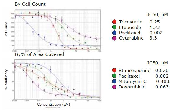
Hela cells were seeded in 96-well plates (4000 cells/well) according to Protocol requirements, and cells were treated with anticancer compounds for 48 hours, and cell images were acquired using the transmission light channel of the MinMax imaging system, and without the need for dyes, only The logic of the software allows you to count cells or derive the percentage of cell coverage. The IC50 value is directly obtained using SoftMax6.4 software, and data can be collected at different time points.
Hepatocyte toxicity test
Hepatocytes differentiated from ipsc (induced pluripotent stem cells) from Cellular Dynamics International (CDI) were used and seeded in microplates as required. Use a collagen-coated 96-well plate with a cell density of 60,000 cells/well, or use a collagen-coated 384-well plate with a cell density of 15,000 cells/well for 2-3 days, then stimulate the cells with the corresponding compound. 72 hours. Cells were stained with Calcein (Viable Cell Assay Kit, Invitrogen) or AF488 (fixed cells, BD kit) dyes conjugated with phalloidin, and images were acquired using the MinMax imaging system. The proliferation of cells.
Hematopoietic stem cell differentiation & toxicity
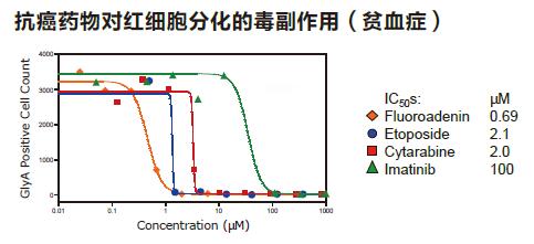
Hematopoietic stem cells (CD34+ cells, Lonza) were seeded in 96-well plates, and then incubated with cytokine indicators and anticancer drugs for 5 days. Cell Tracker Green (Invitrogen) or anti-Glycophorin A (BD kit) was used. After imaging the cells, use the software's own module to analyze the number of cells.
Simplify cell counting in imaging mode
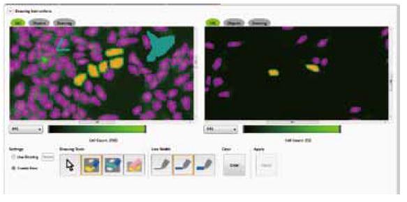
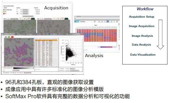
The new SpectraMax MinMax 300 imaging system allows users to easily set up tests to suit their needs and then quickly test them. This system has powerful functions for acquiring and analyzing images. Thanks to the image analysis industry leader software MetaMorph, we integrated its core analysis functions into SoftMax Pro software. Users can select from several application templates based on their specific test needs to acquire and analyze images.
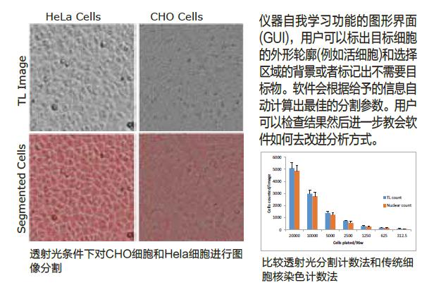
Transmitted light can be segmented according to cell morphology
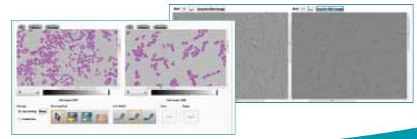
The transmitted light imaging function is very attractive for cytology-based assays because it requires no labeling and requires only a short exposure time, can be detected at different time points and is harmless to cells. However, segmentation of many closely connected cells under transmitted light is a significant challenge because the contrast between the cells and the background is low, which requires the software to have powerful logic. Here, we introduce the patented label-free cell detection technology, which simplifies the analysis of images by the learning ability of the instrument, which can be applied to many different kinds of cells.
Cytology-based assay
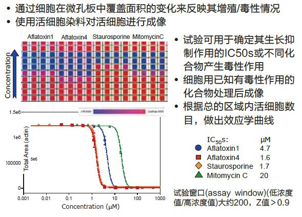
Cell proliferation/toxicity, evaluation of the efficacy of anticancer drugs by the proliferation of cancer cells (HeLa)
The label-free cell counting function under transmitted light conditions can be obtained at different time points by performing cell proliferation assays. It is possible to select the effect of anti-proliferative drugs or growth factors on the cells at different time points, draw the concentration effect curve and give the IC 50 s value, and it is important that the whole process does not need to be stained for observation, and the test process is not Will affect the normal changes of cells.
Hepatocyte toxicity assay using hepatocytes differentiated with ipsc (inducing pluripotent stem cells)
Hepatotoxicity assays were performed using hepatocytes that induced differentiation of pluripotent stem cells (ipsc). After treating these cells for 72 hours with several hepatotoxic drugs, we evaluated the viability of the cells using Calecin or phalloidin-conjugated AF-488. The toxicity assay can be based on changes in cell area or cell number. The IC 50 s values ​​for aflatoxin, erectin, etoposide and other hepatotoxic compounds were determined to have a window of approximately 200 and a Z value of >0.9.
Hematopoietic Stem Cell Differentiation & Toxicity
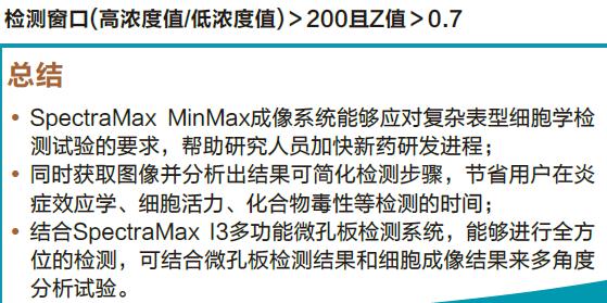
We have designed a test that is very suitable for detecting the effects of growth factors and anticancer drugs on hematopoiesis. We add different growth factors to the hematopoietic stem cells for culture. In addition, we can evaluate the effects of several anticancer drugs, using markers. Antibodies stained with FITC cell lines were stained or cell tracer green dye (Cell Tracker Green) was used. May be used to detect all of the number of cells or cell lineage-specific expression of a number of markers, IC 50 s are determined by different factors. The test window is >100 and the Z value is >0.7.
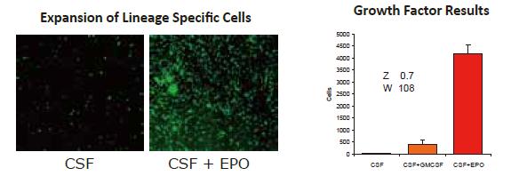
· Effects of different combinations of cytokines on culture of CD34+ cells
· Use cell tracker green dye (Cell Tracker Green) (counting for all cells) or type A glycemic protein (labeled red progenitor cells)
· Add erythropoietin (EPO) and anticancer drugs to CD34+ cells
· Cell count (amplification / toxicity)
Pain Relief Patch For Breast
Pain Relief Patch for Breast
[Name] Medical Cold Patch
[Package Dimension] 10 round pieces
The pain relief patch is composed of three layers, namely, backing lining, middle gel and protective film. It is free from pharmacological, immunological or metabolic ingredients.
[Scope of Application] For cold physiotherapy, closed soft tissue only.
[Indications]
The patches give fast acting pain relief for breast hyperplasia, breast fibroids, mastitis, breast agglomera tion, swollen pain.
[How To Use a Patch]
Please follow the Schematic Diagram. One piece, one time.
The curing effect of each piece can last for 6-8 hours.
[Attention]
Do not apply the patch on the problematic skin, such as wounds, eczema, dermatitis,or in the eyes. People allergic to herbs and the pregnant are advised not to use the medication. If swelling or irritation occurs, please stop using and if any of these effects persist or worsen.notify your doctor or pharmacist promptly. Children using the patch must be supervised by adults.
[Storage Conditions]
Store below 30c in a dry place away from heat and direct sunlight.
Pain Relief Patch For Breast,Pain Relief Plaster For Breast,Relief For Breast Pain,Pad Relief Patch For Breast
Shandong XiJieYiTong International Trade Co.,Ltd. , https://www.xijieyitongpatches.com
