Near-infrared imaging can be used to image deep tissue in mice, including lymph nodes, tumors, and cerebrovascular. The second near-infrared window (1000-1400 nm) fluorescent material has less absorption and scattering of blood and tissue than the first near-infrared window (750-1000 nm) material, and has deeper penetrating ability to living tissue, and is higher in imaging. Signal to noise ratio. Although single-walled carbon nanotubes, rare earth materials, silver sulfide quantum dots, and the like all emit fluorescence in the second near-infrared window, the quantum yield is low. The Takashi Jin research group of the Japan Institute of Physical and Chemical Research has prepared lead sulfide quantum dots, and the quantum yield is obviously improved. The emission of 1100 nm can be obtained by excitation at 785 nm, and the fluorescence intensity is ten times stronger than that of silver sulfide quantum dots; the transmission electron microscope shows that the diameter is less than 10 nm. Very stable in aqueous solution.
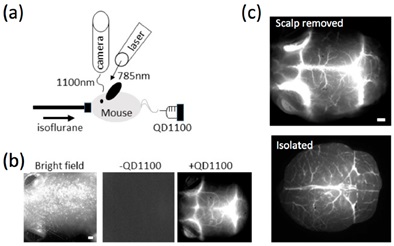
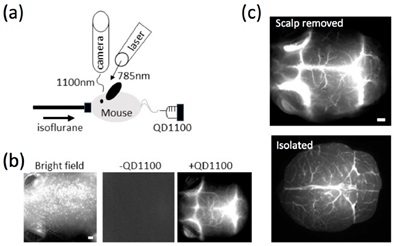
Yukio Imamura et al. injected the lead sulfide quantum dot (QD1100) into the mouse through the tail vein and found that the near-infrared fluorescence signal in the brain of the mouse was clear, clearly showing the structure of the blood vessels in the brain (Fig. 1). A Z-axis stratified scan of the isolated brain tissue was performed to determine that the maximum depth of the QD1100 for cerebrovascular imaging was 1.6 mm. Next, the researchers induced thrombosis of blood vessels in the brain by intraperitoneal injection of LPS. After 18 hours, heparin (hemagglutination inhibitor) was injected into the tail vein, and finally QD1100 was injected intravenously 10 minutes before imaging. Near-infrared imaging clearly showed the pathological state of the brain in mice: LPS stimulated the increase of cerebral vascular plaques, and the addition of heparin reduced the number of plaques by 60% (Fig. 2). For the first time, this study clarified that lead sulfide quantum dots can be used as near-infrared probes to effectively evaluate the pathological state of brain tissue. It is of great significance for the diagnosis of sepsis encephalopathy without damage.

Figure 1. Lead sulfide near-infrared quantum dots (QD1100) show the vascular structure of brain tissue.
(a) QD1100 is injected intravenously into anesthetized mice;
(b) In vivo imaging of brain tissue: brightfield imaging (left), near-infrared imaging of the QD1100 (middle), near-infrared imaging of the QD1100 (right);
(c) Cerebrovascular near-infrared imaging: The image above shows the removal of the scalp, and the image below shows the image after separation.

Figure 2 Near-infrared quantum dot imaging shows cerebral thrombosis in LPS-induced sepsis mice.
(a) Separation of near-infrared imaging of blood vessels in the brain, and the lower row of pictures is an enlargement of the blue frame of the upper row of pictures;
(b) Number of cerebral thrombotic plaques in each group of mice.
Source of the document:
Imamura Y, Yamada S, Tsuboi S, Nakane Y, Tsukasaki Y, Komatsuzaki A, Jin T. Near-Infrared Emitting PbS Quantum Dots for in Vivo Fluorescence Imaging of the Thrombotic State in Septic Mouse Brain. Molecules. 2016;21(8) .
Nuts Seed Kernel include walnut kernel, sunflower seeds kernel, sunflower seed kernel etc., which are excellent food with rich nutrition.
Seed Kernel,Kernel Fruit,Walnut Kernel,Pumpkin seed kernel, sunflower seed kernel
Xi'an Gawen Biotechnology Co., Ltd , https://www.seoagolyn.com