Immunofluorescence technique, also known as fluorescent antibody technology, is one of the earliest developments in marker immunology. It is a technology built on the basis of immunology, biochemistry and microscopy. Some scholars have long tried to combine antibody molecules with some tracer substances, and use antigen-antibody reactions to locate tissue or intracellular antigenic substances.
One. Traditional immunofluorescence experimental steps
(1) Experimental materials
1. Cell samples
2. Reagents, kits: Polymethyl Quan; 70% methanol; C Tong; PBS; Triton; BSA; DAPI dye solution; DABCO; Tris;
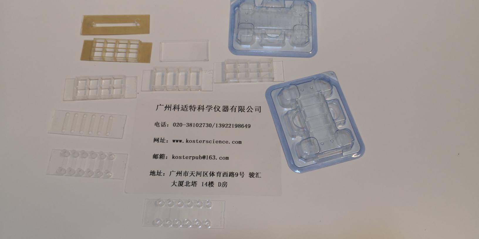
3. Instruments, consumables: slides
(two), experimental steps
Cell slide immunofluorescence experiment steps
first day:
1. Dip the cells that have climbed the cells in the culture plate 3 times with PBS for 3 min each time;
2. Fix the slides with 4% poly-poly- Quan for 15 min, and dip the slides 3 times with PBS for 3 min each time;
3. 0.5% Triton X-100 ( prepared in PBS ) for 20 min at room temperature (the antigen expressed on the cell membrane is omitted);
4. Dip the slides in PBS for 3 times, each time for 3 min, blot the PBS with absorbent paper, add normal goat serum to the slide, and block at room temperature for 30 min;
5. Absorb the blocking solution with absorbent paper, do not wash, add enough amount of diluted primary antibody to each slide and put it into a wet box, incubate at 4 °C overnight;
the next day:
6. Add fluorescent secondary antibody: PBST dip and climb the slide 3 times, each time for 3 min, blot the excess liquid on the slide after the absorbent paper, add the diluted fluorescent secondary antibody, incubate at 20-37 °C for 1 h in the wet box, PBST Dip the slices 3 times for 3 min each time; Note: From the addition of the fluorescent secondary antibody, all subsequent steps are carried out in the darker places.
7. Counterstained nuclei: DAPI was added in the dark for 5 min, the specimens were stained with nuclei, and the excess DAPI was washed out with PBST 5 min×4 times;
8. Absorb the liquid on the climbing plate with absorbent paper, seal it with a sealing liquid containing anti-fluorescence quencher, and observe the captured image under a fluorescence microscope.
The main problems of the traditional immunofluorescence experiment
1. The effect of climbing is not good
2. Large amount of cells and reagents, high cost
3. Uneven distribution of dye liquor
4. Operation is cumbersome, time-consuming and laborious
Introduction to integrated immunofluorescence technology Â
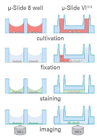
Integrated culture chamber-fixation-staining-imaging immunofluorescence solution 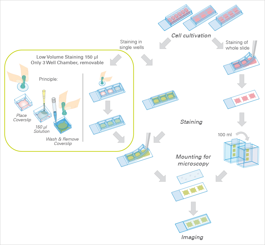
Technical features:
1. Quick and easy sample preparation, integrated culture chamber simplifies immunofluorescence staining
2. Uniform cell distribution, channel slide geometry can produce uniform cell distribution
3. Affordable experimental protocol requires a small amount of cells and reagents
4. High resolution imaging for wide field fluorescence, confocal imaging, FRAP-FRET-FLIM and high quality phase contrast imaging (Phase Contrast)
Â
Main working mode:
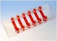
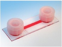
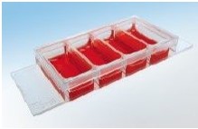
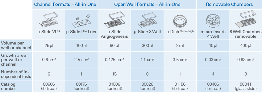
1. Channel mode: ibidi channel slides are ideal for use with small amounts of reagents. μ-Slides VI0.4 is ideal for general immunofluorescence assays, while μ-Slide I0.4Luer allows immunofluorescence staining in low-density culture and flow experiments.
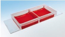
2. Open chamber mode
The Ibidi μ-Slide 8 well and μ-Dish35mm enable standard immunofluorescence protocols in the entire lumen, without coverslips. After staining, the sample was viewed through the bottom of the polymer coverslip using a high resolution microscope.
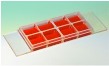
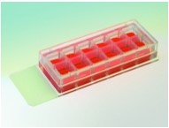
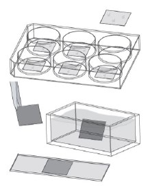
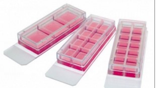
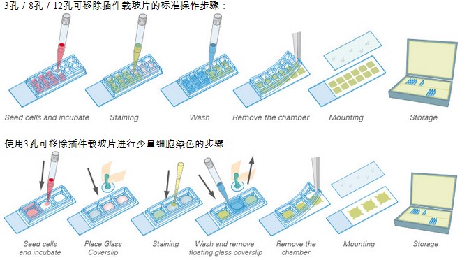
3. Removable Chamber Slide Mode The removable silicon gasket with chamber provides the ideal mode for individual cell culture and incubation. The ibidi micro-Insert 4 Well can be used with very low reagent volumes. Removable 3Well|8Well|12 Well Chambers are ideal for long-term sample storage.
- Integrated immunofluorescence technology application examples
1. Human umbilical vein endothelial cells (HUVEC) cultured under flow conditions in μ-Slide I  0.4  Luer
blue: Â Nucleus (DAPI)
green: Â VE-cadherins (Alexa 488 conjugated antibody)
Red: actin cytoskeleton (Cy5 conjugated antibody)
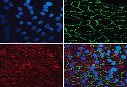
2. Cell line Madin-Darby canine kidney (MDCK) cultured in μ-Slide VI 0.4
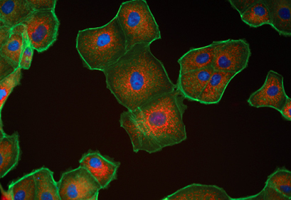
blue: Â Nucleus (DAPI)
green: Â Actin cytoskeleton (Alexa 488 conjugated antibody)
Red: mitochondria (MitoTracker)
3. Differentiated mouse fibroblasts cultured on elastic surface  (ESS 28 kPa)
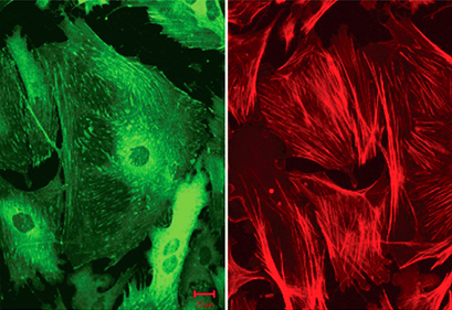
green: Â Zyxin (Alexa 488 conjugated antibody)
Red: alpha-smooth muscle actin (Cy5 conjugated antibody)
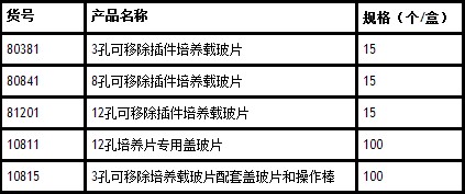
Product technical parameter list
V. Summary and evaluation
(1) This type of chamber slide/culture dish does not require coating, does not require climbing, cultures cell-wash slides-fixed cell-staining-imaging, all steps can be done on this slide. Direct microscopy imaging is effective;
(2) If you want to finish the experiment, you can also seal the film for permanent storage. There are removable cavity slides, 3 holes, 8 holes and 12 holes. According to the experiment, you can tear the cells after dyeing. Drop the frame, observe directly or save the cover;
(3) If the experiment wants to make a small volume, this channel slide is compared with the common immunofluorescence experiment. All the immunofluorescence steps can be completed with the gun adding liquid and aspirating, the operation is simpler; the cell saving reagent is saved, one The channel requires only 25 microliters of liquid; it ensures uniform cell distribution and better observation results;
(4) If the experiment needs to be done in large capacity, or for long-term live cell observation, the IBIDI culture dish has better imaging effect than the ordinary slide glass, and can directly complete all cell operations and observations. This has a matching cover. It can reduce pollution, prevent evaporation, and the lid can be locked without locking. If it is locked, the evaporation rate is basically zero, which is suitable for observing cells for a long time;
(5) If the experiment requires regular observation of the same cell / re-recovering a previously observed cell, or counting cells, there is a petri dish with a counting cell and a chamber slide (8-well), just remember the cell The coordinates of the same cell can be easily retrieved each time, not as accurate as the commonly used marker marker, and the trace of the marker will also affect the observation;
(6) If the experiment is to do high-throughput immunofluorescence, such as drug sieve, there are 24-well plates, 96-well plates, 384 wells, the price is similar to the porous plate in the market, the porous plate does not need to be coated, and there is no spontaneous Fluorescence, the pore walls are black, so the immunofluorescence between the pores and the pores does not affect each other;
(7) These consumables are specifically designed for confocal super-resolution microscopy imaging with good results;
(8) These products are processed by ibidi's patented physical method ibiditreat, the bottom is ultra-thin and hydrophilic, most types of cells can be directly attached to the wall, no additional coating is required; if there are special experimental needs, You can repack it yourself (choose the uncoated version);

herpes zoster vaccine,Hepatitis B Vaccine For Adults,Tetanus Booster,Td Vaccine
FOSHAN PHARMA CO., LTD. , https://www.full-pharma.com